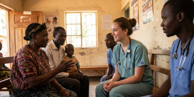
Clinical observation in resource-limited East African settings places you in environments where physical examination does the work that laboratory tests do elsewhere. Before you can learn from what you observe, you need to know what you are looking at. Let’s cover the most common conditions you will encounter, what they look like, and how to build the pattern recognition that turns observation into genuine clinical learning.
Key Highlights
- Visual pattern recognition is a primary diagnostic tool in resource-limited East African clinical settings where laboratory access is constrained.
- Malaria, severe anemia, malnutrition, and typhoid each present with specific, observable signs that clinical staff assess in seconds.
- Conjunctival pallor is the most reliable visual indicator of anemia across all skin tones and requires no equipment.
- Kwashiorkor and marasmus present completely differently despite both being forms of severe malnutrition, and learning to distinguish them visually sharpens clinical observation.
- The pre-encounter observation practice – five to ten seconds of independent visual assessment before each clinical encounter – is the most effective way to develop pattern recognition during a placement.
- Your observations belong in your reflective journal, not in clinical conversations or recommendations to patients.
Why Visual Assessment Carries More Weight in Resource-Limited Settings
In a well-resourced North American clinical environment, a physician who suspects malaria orders a rapid diagnostic test. A physician who suspects anemia orders a complete blood count. The physical examination findings that prompted those orders are noted but rarely function as the primary diagnostic tool. Research on clinical management in resource-limited settings documents that experienced clinicians in these environments derive a substantially higher proportion of their diagnostic information from direct physical observation precisely because laboratory backup is unavailable or delayed.
Observing clinical practice in East African settings gives you access to physical examination used in its most fundamental form: as the primary instrument of clinical reasoning rather than as the prompt for a laboratory order. Watching a clinical officer assess a febrile patient with suspected malaria using only their eyes, hands, and clinical judgment makes the relationship between physical signs and clinical conclusions visible in a way that technology-dependent practice often obscures.
The WHO Integrated Management of Childhood Illness framework was developed for clinical environments where laboratory support is limited. It provides the structured visual assessment approach used by clinical staff across East African settings. Familiarizing yourself with the IMCI visual assessment criteria before your placement gives you the framework to understand what you are watching when clinical staff assess pediatric patients.
Malaria: The Most Common Febrile Illness in the Region
What You Will See
Malaria caused by Plasmodium falciparum is the dominant febrile illness in most East African outpatient settings, and experienced clinicians develop a rapid visual sense of the malaria patient before the rapid diagnostic test confirms it. The presenting patient is typically febrile, which you may observe as flushed skin, sweating, or in severe cases a visibly altered level of consciousness. Rigors – episodes of intense shaking and shivering – are characteristic of the cyclic pattern of malarial fever and can be dramatic when you observe them for the first time.
In children with severe malaria, look for pallor of the conjunctiva and palmar creases, indicating hemolytic anemia. Jaundice – a yellowing of the skin and whites of the eyes – indicates hemolysis and is visible in moderate to severe cases without any laboratory support. Prostration, the inability of a child to sit upright unassisted, is a WHO danger sign for severe malaria and is assessed visually at the start of every pediatric encounter.
What the Clinical Team Is Doing
Watch how the clinician approaches the febrile child. They are typically assessing level of consciousness first, then the ability to sit or stand, then the conjunctiva and palmar creases for pallor, then the skin and eyes for jaundice, and then the abdomen for splenomegaly – the enlarged spleen that develops with repeated malaria infections. Each assessment takes seconds and generates clinical information that shapes the severity classification and treatment decision before any test result is available.
Severe Anemia: Visible in the Conjunctiva and Beyond
Severe anemia is among the most prevalent serious conditions in East African pediatric and maternal populations, driven by malaria, iron deficiency, hookworm, and HIV. It is also one of the conditions most reliably assessed through visual examination.
The conjunctival assessment is the most reliable visual sign across diverse skin tones: pull down the lower eyelid to expose the inner surface and look at its color. Pink and well-perfused is normal. Pale or white indicates significant anemia. Palmar crease pallor – visible even in patients with dark skin when the hand is held open, and the palm is stretched slightly – is the second most reliable visual sign. Severe pallor of the lips and oral mucosa indicates very significant anemia. Koilonychia, the spoon-shaped nail deformity of chronic iron deficiency, is visible to a prepared observer who knows to look for it.
Beyond the direct pallor signs, watch for behavioral and functional indicators: the child who tires quickly, the adolescent girl who is breathless at rest, the pregnant woman whose fatigue seems disproportionate to her presentation. These functional signs of inadequate oxygen delivery are part of what the experienced clinician is reading when they observe a patient before the formal examination begins.
Malnutrition: Two Distinct Visual Presentations
Marasmus
Marasmus results from severe total caloric deficiency. The child with marasmus has lost both fat and muscle mass and presents with a characteristic wasted appearance: visible ribs and vertebrae, loose skin hanging in folds over the upper arms and thighs, prominent bony landmarks at the shoulders and hips, and a facial appearance that has lost the rounded contours of normal childhood. The marasmic child is typically alert, often visibly distressed or irritable, and hungry. No edema is present.
Kwashiorkor
Kwashiorkor results from severe protein deficiency with relatively preserved caloric intake and presents strikingly differently. The child with kwashiorkor may appear deceptively plump due to generalized edema, most evident in the feet and lower legs. The hair changes are among the most visually distinctive signs in global pediatric medicine: hair that was previously dark and tightly curled becomes reddish, brownish, or even blonde in severely affected children. The skin develops depigmented patches and, in severe cases, cracks and peels. The child with kwashiorkor is typically listless and anorexic, in direct contrast to the alert and hungry marasmic child.
Scabies and Skin Infections: High Volume in Outpatient Settings
Skin conditions account for a substantial proportion of outpatient presentations in many East African settings, and several of the most common are visually distinctive once you have seen them. Scabies presents with an intensely itchy papular rash concentrated in characteristic locations: the webspaces between fingers, the wrists, the waist, and the ankles. The burrow tracks left by the mite are visible as tiny linear marks in the skin. Secondary bacterial infection from scratching is common and produces impetigo-like crusting over the primary rash.
Tinea capitis – ringworm of the scalp – presents as patchy hair loss with scaling and sometimes a kerion, a raised, boggy, inflammatory mass that can look alarming to an unprepared observer. Tropical ulcers, chronic skin ulcers typically on the lower leg, are commonly seen in outpatient settings and can be large even when not acutely infected.
How to Practice Visual Recognition as an Observer
The most effective practice is a simple discipline: before each patient encounter, you observe and take 5 to 10 seconds to form your own visual impression. Note their level of alertness, their posture, their skin color as visible from a distance, their apparent level of distress, and any immediately visible physical signs. Do not share this impression. Form it, then watch the clinical examination unfold and compare what you saw to what the clinician finds.
Over many encounters, this practice builds the pattern-recognition skills that underlie clinical reasoning. Your early impressions will be vague and often inaccurate. By the end of a two-week placement, you will notice your impressions becoming more specific and more frequently consistent with the clinical findings that follow.
Students who want to understand how these observational skills translate into application value should read about what clinical experience demonstrates to admissions committees. The specific, concrete clinical observations you develop the vocabulary to describe during international placements are exactly what distinguishes a compelling clinical narrative from a generic one.
Common Mistakes When Developing Visual Recognition
The most common mistake is attempting to share visual impressions with clinical staff or patients during the shift. Your role is to observe and learn, not to contribute clinical opinions. A student who tells a nurse, “I think this patient looks anemic,” is overstepping, regardless of whether the observation is accurate.
A second mistake is over-generalizing from limited exposure. Seeing one presentation of kwashiorkor does not make you competent to recognize all presentations. Clinical pattern recognition requires repeated exposure across varied presentations, and the appropriate response to growing observational skill is curiosity and continued learning rather than premature confidence.
A third mistake is allowing the visual intensity of clinical presentations in resource-limited settings to shift from educational to voyeuristic. You are observing because it makes you a better clinician, not because the presentations are dramatic.
Connecting This to Global Health and Your Future Training
The conditions described in this article are common across the East African facilities where IMA places students. Understanding what global health observation builds in a pre-health student includes this kind of clinical pattern recognition that transfers directly into medical school physical examination training. Students who arrive at medical school with developed observational habits and clinical vocabulary have a meaningful advantage in the physical examination curriculum.
Students who are also reflecting on their shadowing hours and how to frame international clinical exposure on their applications should read about what makes the shadowing experience meaningful to admissions committees.
Frequently Asked Questions
How long does it take to develop reliable visual recognition of these conditions?
Pattern recognition builds across many exposures rather than from a few. Most students notice their impressions becoming meaningfully more accurate after two to three weeks of consistent structured observation. Full clinical competency in visual diagnosis requires years of supervised practice. The goal during a pre-health placement is foundational vocabulary and pattern recognition.
Should I mention conditions I think I recognize to clinical staff?
No. Your role is to observe. The appropriate channel for observations you develop is your reflective journal and, during appropriate pauses in clinical activity, specific questions to your supervisor about what you observed. Not during active patient encounters.
Does skin tone affect my ability to see pallor signs?
Some pallor signs are affected by skin tone and some are not. Conjunctival pallor is reliable across all skin tones because the conjunctival mucosa has consistent baseline color regardless of skin pigmentation. Palmar crease pallor is also useful across diverse skin tones when the hand is opened and slightly stretched. Lip and facial skin pallor are less reliable with darker skin tones. Experienced clinicians in these settings use conjunctival and palmar assessment as their primary visual tools for this reason.
What if I notice a visual sign that the clinical staff has not commented on?
Note it in your journal after the shift and ask your supervisor about it during a non-clinical moment. It is possible you noticed something significant. It is also possible you misread a normal variant. Ask rather than act.
Is it appropriate to ask a clinician to explain what they are looking for during an assessment?
Yes, during appropriate moments. Not during the patient encounter itself. During a natural pause between patients or after rounds, a brief specific question is almost always welcome. What were you assessing when you pulled down the child’s eyelid is exactly the kind of question that demonstrates genuine engagement.
How do I document what I observe without violating patient privacy?
Document the clinical presentation and visual signs in de-identified terms. The condition and visual findings are appropriate content. Names, specific ages combined with diagnoses, room numbers, and any identifying detail are not. A journal entry describing a child of approximately two years with classic kwashiorkor including hair color change and pedal edema is appropriate. The same entry with a name or ward number is not.
Will these observational skills be relevant once I am in medical school?
Yes. Physical examination skills are foundational to clinical medicine regardless of resource context. Students who arrive at medical school with developed observational habits and clinical pattern recognition have a vocabulary for the physical examination curriculum that their classmates are building from scratch.
Are conditions in outpatient settings different from what I will see in inpatient wards?
The distribution differs. Outpatient settings see a high volume of malaria, upper respiratory infections, skin conditions, and early presentations of typhoid and malnutrition. Inpatient wards see more severe presentations. Pediatric wards have the highest concentration of IMCI-relevant presentations. Knowing your setting helps you anticipate which conditions to prepare for.