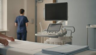
Medical Ultrasound Awareness Month runs every October, and the Society of Diagnostic Medical Sonography has promoted it since 1992. For students considering imaging careers, it is a useful prompt to study how sonographers, radiologic technologists and radiologists divide the work, and to plan observation that shows you more than a single scan.
A career in imaging can look like a list of machines. Spending time with the people who run them tells you much more. If you are early in a pre-rad tech path, IMA’s guide to radiologists and radiographers explains the basic division of roles before you watch it in practice.
Use This Year’s Campaign to Sharpen Your Questions
The SDMS describes Medical Ultrasound Awareness Month as a month held annually in October to increase awareness of diagnostic medical sonography. It was previously known as Sonography Awareness Week and then Sonography Awareness Month, and the society’s theme for 2026 is “Inside Images.”
That theme works as a student exercise. Every image you see in a department has a story behind it: why the exam was ordered, how the patient was prepared, which technique was chosen, how quality was checked and who interpreted the result. Pick one part of that sequence before each observation day and follow it closely.
What Does a Sonographer Actually Do?
The Bureau of Labor Statistics explains that diagnostic medical sonographers operate equipment that creates images of the inside of the body. Many specialize. Abdominal sonographers image organs such as the kidney, liver and pancreas, cardiac sonographers image the heart, vascular sonographers evaluate blood vessels and blood flow, and obstetric and gynecologic sonographers image the female reproductive system.
Ultrasound uses sound rather than X-rays. The National Institute of Biomedical Imaging and Bioengineering notes that diagnostic ultrasound does not produce ionizing radiation like that produced by X-rays, and that monitoring the growth of a fetus during pregnancy is one of its most common uses. The same page adds that professional groups recommend using it only when there is a true medical need.
How Do Sonography and Radiologic Technology Compare?
The two fields share a department but differ in equipment, training and day-to-day work. The figures below come from the Bureau of Labor Statistics Occupational Outlook pages for sonographers and for radiologic and MRI technologists, using 2025 data and 2025 to 2035 projections.
| Measure | Diagnostic medical sonographers | Radiologic and MRI technologists |
|---|---|---|
| Median annual wage, May 2025 | $96,590 | $81,390 |
| Jobs in 2025 | About 92,200 | About 278,100 |
| Projected growth, 2025 to 2035 | 14 percent | 5 percent |
| Projected openings each year | About 6,000 | About 15,300 |
| Typical entry-level education | Associate’s degree or postsecondary certificate | Associate’s degree |
These numbers describe national labor markets. They do not tell you which role you will enjoy, which program will accept you or what any single employer pays. IMA’s sonographer salary guide and radiologic technologist salary guide go further into pay by setting and state.
Watch the Whole Imaging Team
An imaging exam involves more people than the person at the controls. Someone receives the request, someone prepares and positions the patient, someone checks that the images are usable and a physician interprets them. Each handoff is a place where information can be clarified or lost.
The table below is an original observation template. Use it within your host’s rules, and leave names, dates of birth and other identifiers out of your notes.
| Moment | What to observe | Question for your supervisor |
|---|---|---|
| Before the exam | How the request is checked and the patient is identified | What do you confirm before starting? |
| Preparation | How the procedure is explained and consent is handled | How do you adjust an explanation for a worried patient? |
| Positioning and technique | How the technologist chooses angles and settings | What makes you repeat an image? |
| Safety | Where staff stand and how shielding or distance is used | Which safety step do students most often overlook? |
| Quality check | How images are reviewed before the patient leaves | What counts as a diagnostic image here? |
| Interpretation | How the technologist and radiologist communicate | What information helps the radiologist most? |
You will not get through every row in one day. Choose two, ask when there is a quiet moment and write down the answer in your own words afterward.
Why Does Patient Communication Matter So Much in Imaging?
Many imaging exams ask patients to hold still, hold their breath, change position or wait alone in a room with large equipment. How a technologist explains those requests affects both the patient’s experience and the quality of the image. Listen to the exact words staff use, notice how they check understanding and pay attention to what happens when a patient is in pain or anxious.
This is one of the most transferable things you can learn. Whatever imaging career you choose, you will spend much of each day explaining short, specific instructions to people who may be frightened.
Turn One Exam Into a Useful Reflection
Consider a fictional teaching example. A student watches a technologist repeat a chest X-ray after reviewing the first image. A basic note reads, “I watched a chest X-ray.” A stronger reflection reads, “I noticed that the technologist checked the first image before the patient left and chose to repeat it. When I asked why, she explained what made the first image less useful. I had assumed that once an image was taken, the work was done.”
The second version shows what you learned and what changed in your thinking. It does not claim that you judged the image yourself or that you know the clinical result. Use the same structure for your own notes: what you saw, what the professional explained and what you still want to understand.
If you observe in a hospital where staff speak a language you are still learning, clarify what you heard before you interpret it. IMA’s guide to staying engaged during observation across a language barrier offers practical ways to keep learning when you miss parts of a conversation.
Keep Your Role Clear
Observing an imaging department does not make you responsible for operating equipment, positioning patients, reading images or sharing results. Ask what students are permitted to do before the day starts, follow every radiation safety instruction, including where to stand during an exam, and remember that a patient can decline to have a student present.
The same honesty belongs in your applications. Describe observation as observation. If you hope to count hours toward a program, ask that program in writing which settings and supervisors it accepts and how it wants the experience documented. Sonography and radiography credentials are earned through accredited education and certification in your own country, not through time abroad.
Choose an Experience That Answers Your Questions
After a few days of observation, write down one question you could not answer. Perhaps you understand X-ray workflow but not how a department decides between imaging methods, or you have seen routine exams but not how urgent cases are prioritized. That gap is a good guide for choosing your next experience.
IMA’s radiology internships abroad place pre-rad tech and radiologic technology students in the radiology department of Coast General Teaching and Referral Hospital in Mombasa, Kenya, for stays from two weeks, usually six to eight hours a weekday. Licensed clinicians supervise interns, and staff are on call 24/7. The program page lists X-ray, CT and MRI among the imaging interns see, and what you see depends on the equipment in service, the day’s cases and your level of training.
Ultrasound is also central to pregnancy care, so students curious about obstetric imaging may find it useful to read how National Midwifery Week frames observation on a maternity service.
A good observation day leaves you with a clearer picture of the team and a sharper question for next time. That is a stronger outcome than a long list of machines you watched someone else operate.
Frequently Asked Questions
When Is Medical Ultrasound Awareness Month?
It is held every October. The Society of Diagnostic Medical Sonography has promoted it since 1992, and its 2026 theme is “Inside Images.”
What Is the Difference Between a Sonographer and a Radiologic Technologist?
Sonographers create images with ultrasound, which uses sound waves. Radiologic technologists perform diagnostic imaging exams such as X-rays, and MRI technologists operate MRI scanners. Training, equipment and daily work differ between the roles.
Will I Observe Ultrasound During an IMA Radiology Internship?
Do not assume so. IMA’s program page lists X-ray, CT and MRI among the imaging interns see, and what you observe depends on the equipment in service, the cases that day and your level of training.
Can I Operate Imaging Equipment as a Student Observer?
No. Pre-rad tech students mainly observe. Staff decide whether you may help with any simple, non-invasive task, and you never read images or give results.
Does Observation Abroad Count Toward Imaging Program Requirements?
Each program sets its own rules. Ask the programs you plan to apply to which settings and supervisors they accept, and how they want hours documented, before relying on them.
What Should I Write After an Imaging Observation?
Record one specific moment, what the technologist or radiologist explained and what remains unclear to you. Leave out patient identifiers and avoid claims about diagnoses or results you did not follow.